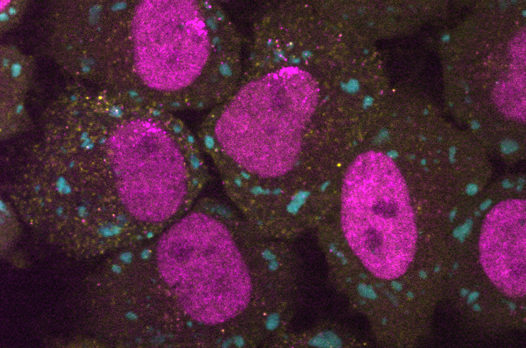
Spotting the Unexpected: Translation in Stress Granules

When cells are exposed to diverse stress stimuli (e.g., viral infection, heat or oxidative stress), they activate the integrated stress response (ISR) pathway to promote cellular recovery. This pathway results in a decrease of global protein synthesis, an induction of selected genes and the assembly of cytoplasmic membrane-less organelles termed stress granules (SGs). For a long time, it was widely believed that SGs promote the translational repression of RNAs and that they contain exclusively translationally-repressed RNAs. A recent single-molecule imaging study from Jeffrey Chao’s lab at the FMI Basel now challenges this notion (Mateju et al., Cell, 2020).
To characterize the relationship between mRNA localization and translation during stress, Mateju and colleagues aimed to simultaneously image individual mRNA molecules and the associated nascent polypeptide chains at defined sub-cellular localizations. To do so, they engineered a reporter construct containing the 5’ UTR of ATF4 (a gene that is induced upon stress conditions), followed by a coding sequence encoding a SunTag array and MS2 stem-loops in the 3’ UTR. Upon integration and expression of this reporter construct in HeLa cells, the individual reporter mRNAs and the nascent peptide chains were visualized in single-molecule imaging experiments via the MS2 stem-loops and the SunTag, respectively. Combining these experiments with live-cell imaging of SGs under oxidative stress conditions enabled the analysis of translation activity of the reporter mRNAs both in the cytoplasm and SGs in stressed cells.
As expected, the authors observed many non-translated mRNAs (i.e., mRNAs devoid of a SunTag signal) in SGs. But surprisingly, ATF4-SunTag mRNAs undergoing translation are also found in SGs. In fact, about 30% of ATF4-SunTag mRNAs localized to SGs are translated, suggesting that SG-associated translation is not a rare event for this type of transcript. “I was very surprised to see translation activity in SGs. Initially, we were skeptical that SG-localized mRNAs could be translated efficiently, but with further experiments and quantitative analysis we became more convinced.”, says Daniel Mateju, first author of the study.
To exclude the possibility that the mRNAs with nascent polypeptides in SGs are aberrant or stalled translation complexes, Mateju and colleagues sought to further characterize SG-associated translation. First, by acquiring longer movies, they identified SG-localized mRNAs initially devoid of SunTag signal but acquired SunTag signal over time, indicating that translation initiation can take place in SGs. Second, the authors quantified elongation rates and found that the elongation rates of SG-localized and cytoplasmic mRNAs are comparable. And third, by assessing the kinetics of SunTag signal accumulation of SG-localized transcripts in time-lapse movies, they concluded that ribosomes are able to terminate translation in SGs. Thus, mRNAs can undergo the entire translation cycle (initiation, elongation and termination) in SGs and SG-associated translation resembles translation in the cytosol.
The authors further obtained evidence that SG-associated translation is not limited to stress response mRNAs such as ATF4. When the 5’UTR was replaced with one containing a 5’TOP motif (a cis-acting motif found in genes that are repressed during stress), this reporter mRNA was strongly translationally repressed. Only a few 5’TOP reporter mRNAs were found to be translating during stress and those were found not only in the cytosol but also in SGs. Taken together, these results show that mRNAs localized to SGs can undergo translation and suggest that the SG environment per se does not induce translation repression.
The recent technological developments in single-molecule analysis have revolutionized the way the function of membrane-less organelles or biomolecular condensates can be dissected and open new avenues for future research. Jeffrey Chao, corresponding author of the study, is excited about this development: “For a long time, we could only observe stress granules and look at the juxtaposition of stress granules and P-bodies. Now we can start testing the models that have been proposed and find out which ones are correct and which need to be re-evaluated.”
Mateju et al. (2020) Cell, 183(7), 1801-1812.e13
Text: Veronika Herzog