Disrupting Telomerase
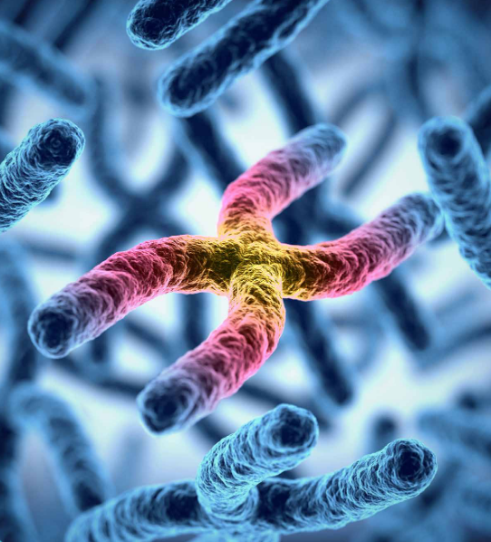
How to stop cancer cells from proliferating unlimitedly? That is one of the central questions in cancer research, as these cells have found several ways to circumvent normal cell aging. One of the key players in this is telomerase, which is highly active in cancer cells, keeping their telomeres intact and making the cells virtually immortal. Telomerase counteracts telomere shortening and cellular senescence in germ, stem, and cancer cells by adding repetitive DNA sequences to the ends of chromosomes. In normal cells telomeres are susceptible to damage by reactive oxygen species (ROS), but the exact consequences of oxidation of telomeres on telomere length as well as the mechanisms that protect from ROS-mediated telomere damage are not well understood.
Wareed Ahmed and Joachim Lingner from the Swiss Institute for Experimental Cancer Research at the Ecole Polytechnique Fédérale de Lausanne have recently unveiled a new detail in the mechanism of the protection of telomeres. 8-oxoguanine nucleotides at 3’ ends of telomeric substrates efficiently inhibit telomerase in vitro, whereas, at internal positions, they suppress G-quadruplex formation and were therefore proposed to promote telomerase activity. As Ahmed and Lingner show in a recent Genes & Development paper, two cooperating enzymes, peroxiredoxin 1 (PRDX1) and 7,8-dihydro-8-oxoguanine triphosphatase (MTH1) prevent accumulation of oxidized guanine in the genome. Disrupting the peroxiredoxin 1 (PRDX1) and 7,8dihydro8oxoguanine triphosphatase (MTH1) genes in cancer cells led to telemores being susceptible to oxidative stress and damage. As a consequence, the cells’ telomeres shrunk with every round of cell division, eventually disappearing altogether.
By identifying these two enzymes that apparently protect chromosomes from oxidative damage and shortening, the EPFL scientists have uncovered a potential new anticancer strategy for stopping telomerase, the enzyme that immortalizes tumors. So far, attempts to efficiently block telomerase in cancer have not been fruitful in the clinic. The discovery of the cooperating enzymes opens up a new opportunity to indirectly block telomerase. “Instead of inhibiting the enzyme itself, we target its substrate – the chromosome end – making it un-extendable by telomerase,” says Lingner.
To determine the roles of MTH1 and PRDX1 in suppressing oxidation of guanine in the genome, the team stained nuclei of HCT116 wildtype, PRDX1 knockout, MTH1 knockout, and PRDX1/MTH1 doubleknockout cells with antibodies recognizing 8oxoguanine (8-oxo G). It could be shown that 8-oxo G was incorporated into the genome in a oxygen concentrationdependent manner. The staining for 8-oxo G was enhanced in MTH1 knockout cells and further pronounced in the PRDX1/MTH1 double-knockout cells.
The work opens novel avenues to target telomeres and telomerase in cancer cells. Notably, recent data show that cancer cells to be more vulnerable than noncancer cells to ROS. Thus, increasing ROS may preferentially target cancer cells, and several chemotherapeutic cancer drugs as well as ionizing radiation either induce ROS or reduce the cellular antioxidant capacity. The authors ask the intriguing question whether these drugs may be used to target telomeres and whether they can be boosted for telomerase inhibition by combing them with inhibitors for MTH1 and PRDX1.
Ahmed W. and Lingner J. (2018) Genes & Development 32(9–10). 658-669
Text by Roland Fischer